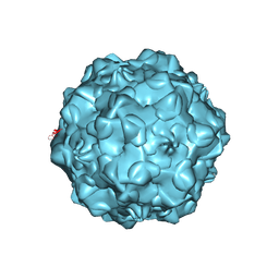
Loading
PDBj
MenuPDBj@FacebookPDBj@X(formerly Twitter)PDBj@BlueSkyPDBj@YouTubewwPDB FoundationwwPDBDonate
RCSB PDBPDBeBMRBAdv. SearchSearch help
PDB: 333 results

4SBV
DownloadVisualize
BU of 4sbv by Molmil
The REFINEMENT OF SOUTHERN BEAN MOSAIC VIRUS IN RECIPROCAL SPACE
Descriptor: CALCIUM ION, SOUTHERN BEAN MOSAIC VIRUS COAT PROTEIN
Authors:Rossmann, M.G.
Deposit date:1985-04-01
Release date:1985-07-17
Last modified:2024-10-23
Method:X-RAY DIFFRACTION (2.8 Å)
Cite:Refined structure of southern bean mosaic virus at 2.9 A resolution.
J.Mol.Biol., 197, 1987
2MEV
DownloadVisualize
BU of 2mev by Molmil
STRUCTURAL REFINEMENT AND ANALYSIS OF MENGO VIRUS
Descriptor: MENGO VIRUS COAT PROTEIN (SUBUNIT VP1), MENGO VIRUS COAT PROTEIN (SUBUNIT VP2), MENGO VIRUS COAT PROTEIN (SUBUNIT VP3), ...
Authors:Rossmann, M.G.
Deposit date:1989-04-21
Release date:1990-01-15
Last modified:2024-11-20
Method:X-RAY DIFFRACTION (3 Å)
Cite:Structural refinement and analysis of Mengo virus.
J.Mol.Biol., 211, 1990
5VU2
DownloadVisualize
BU of 5vu2 by Molmil
Electron cryo-microscopy of "immature" Chikungunya VLP
Descriptor: E1 envelope glycoprotein, E2 envelope glycoprotein, E3 envelope glycoprotein, ...
Authors:Rossmann, M.G, Yap, M.L.
Deposit date:2017-05-18
Release date:2017-12-06
Last modified:2024-10-16
Method:ELECTRON MICROSCOPY (6.8 Å)
Cite:Structural studies of Chikungunya virus maturation.
Proc. Natl. Acad. Sci. U.S.A., 114, 2017
6ODF
DownloadVisualize
BU of 6odf by Molmil
EEEV glycoproteins bound with heparan sulfate
Descriptor: 2-O-sulfo-beta-L-galactopyranuronic acid-(1-4)-[(2R,3R,4R,5R,6R)-2,4,5-trihydroxy-6-(sulfooxy)tetrahydro-2H-pyran-3-yl]sulfamic acid, E1, E2
Authors:Rossmann, M.G, Chen, C.L.
Deposit date:2019-03-26
Release date:2020-04-01
Last modified:2024-11-20
Method:ELECTRON MICROSCOPY (5.8 Å)
Cite:Cryo-EM structure of eastern equine encephalitis virus in complex with heparan sulfate analogues.
Proc.Natl.Acad.Sci.USA, 117, 2020
6NN3
DownloadVisualize
BU of 6nn3 by Molmil
Structure of parvovirus B19 decorated with Fab molecules from a human antibody
Descriptor: Fab monoclonal antibody 860-55D, heavy chain, light chain, ...
Authors:Rossmann, M.G, Sun, Y, Klose, T, Liu, Y.
Deposit date:2019-01-14
Release date:2019-02-13
Last modified:2025-05-14
Method:ELECTRON MICROSCOPY (3.22 Å)
Cite:Structure of Parvovirus B19 Decorated by Fabs from a Human Antibody.
J. Virol., 93, 2019
1GFF
DownloadVisualize
BU of 1gff by Molmil
THE ATOMIC STRUCTURE OF THE DEGRADED PROCAPSID PARTICLE OF THE BACTERIOPHAGE G4: INDUCED STRUCTURAL CHANGES IN THE PRESENCE OF CALCIUM IONS AND FUNCTIONAL IMPLICATIONS
Descriptor: BACTERIOPHAGE G4 CAPSID PROTEINS GPF, GPG, GPJ
Authors:Rossmann, M.G.
Deposit date:1995-11-06
Release date:1996-04-03
Last modified:2024-05-22
Method:X-RAY DIFFRACTION (3 Å)
Cite:Atomic structure of the degraded procapsid particle of the bacteriophage G4: induced structural changes in the presence of calcium ions and functional implications.
J.Mol.Biol., 256, 1996
1JEW
DownloadVisualize
BU of 1jew by Molmil
CRYO-EM STRUCTURE OF COXSACKIEVIRUS B3(M STRAIN) WITH ITS CELLULAR RECEPTOR, COXSACKIEVIRUS AND ADENOVIRUS RECEPTOR (CAR).
Descriptor: COXSACKIEVIRUS AND ADENOVIRUS RECEPTOR, COXSACKIEVIRUS CAPSID, COAT PROTEIN VP1, ...
Authors:Rossmann, M.G, He, Y.
Deposit date:2001-06-19
Release date:2001-10-03
Last modified:2024-02-07
Method:ELECTRON MICROSCOPY (22 Å)
Cite:Interaction of coxsackievirus B3 with the full length coxsackievirus-adenovirus receptor.
Nat.Struct.Biol., 8, 2001
1MEC
DownloadVisualize
BU of 1mec by Molmil
CONFORMATIONAL VARIABILITY OF A PICORNAVIRUS CAPSID: PH-DEPENDENT STRUCTURAL CHANGES OF MENGO VIRUS RELATED TO ITS HOST RECEPTOR ATTACHMENT SITE AND DISASSEMBLY
Descriptor: MENGO VIRUS COAT PROTEIN (SUBUNIT VP1), MENGO VIRUS COAT PROTEIN (SUBUNIT VP2), PHOSPHATE ION
Authors:Rossmann, M.G.
Deposit date:1992-01-17
Release date:1994-01-31
Last modified:2024-10-23
Method:X-RAY DIFFRACTION (3.2 Å)
Cite:Conformational variability of a picornavirus capsid: pH-dependent structural changes of Mengo virus related to its host receptor attachment site and disassembly.
Virology, 175, 1990
1C8E
DownloadVisualize
BU of 1c8e by Molmil
FELINE PANLEUKOPENIA VIRUS EMPTY CAPSID STRUCTURE
Descriptor: FELINE PANLEUKOPENIA VIRUS CAPSID
Authors:Rossmann, M.G, Simpson, A.A.
Deposit date:2000-05-05
Release date:2000-08-09
Last modified:2024-11-13
Method:X-RAY DIFFRACTION (3 Å)
Cite:Host range and variability of calcium binding by surface loops in the capsids of canine and feline parvoviruses.
J.Mol.Biol., 300, 2000
1C8F
DownloadVisualize
BU of 1c8f by Molmil
FELINE PANLEUKOPENIA VIRUS EMPTY CAPSID STRUCTURE
Descriptor: CALCIUM ION, FELINE PANLEUKOPENIA VIRUS CAPSID
Authors:Rossmann, M.G, Simpson, A.A.
Deposit date:2000-05-05
Release date:2000-08-09
Last modified:2024-10-30
Method:X-RAY DIFFRACTION (3 Å)
Cite:Host range and variability of calcium binding by surface loops in the capsids of canine and feline parvoviruses.
J.Mol.Biol., 300, 2000
1C8G
DownloadVisualize
BU of 1c8g by Molmil
FELINE PANLEUKOPENIA VIRUS EMPTY CAPSID STRUCTURE
Descriptor: CALCIUM ION, FELINE PANLEUKOPENIA VIRUS CAPSID
Authors:Rossmann, M.G, Simpson, A.A.
Deposit date:2000-05-05
Release date:2000-08-09
Last modified:2024-10-30
Method:X-RAY DIFFRACTION (3 Å)
Cite:Host range and variability of calcium binding by surface loops in the capsids of canine and feline parvoviruses.
J.Mol.Biol., 300, 2000
1C8D
DownloadVisualize
BU of 1c8d by Molmil
CANINE PANLEUKOPENIA VIRUS EMPTY CAPSID STRUCTURE
Descriptor: CALCIUM ION, CANINE PARVOVIRUS CAPSID
Authors:Rossmann, M.G, Simpson, A.A.
Deposit date:2000-05-05
Release date:2000-08-09
Last modified:2024-11-20
Method:X-RAY DIFFRACTION (3 Å)
Cite:Host range and variability of calcium binding by surface loops in the capsids of canine and feline parvoviruses.
J.Mol.Biol., 300, 2000
1C8H
DownloadVisualize
BU of 1c8h by Molmil
CANINE PARVOVIRUS STRAIN D EMPTY CAPSID STRUCTURE AT PH 5.5
Descriptor: CALCIUM ION, CANINE PARVOVIRUS CAPSID
Authors:Rossmann, M.G, Simpson, A.A.
Deposit date:2000-05-05
Release date:2000-08-09
Last modified:2024-10-09
Method:X-RAY DIFFRACTION (3.5 Å)
Cite:Host range and variability of calcium binding by surface loops in the capsids of canine and feline parvoviruses.
J.Mol.Biol., 300, 2000
1CD3
DownloadVisualize
BU of 1cd3 by Molmil
PROCAPSID OF BACTERIOPHAGE PHIX174
Descriptor: PROTEIN (CAPSID PROTEIN GPF), PROTEIN (SCAFFOLDING PROTEIN GPB), PROTEIN (SCAFFOLDING PROTEIN GPD), ...
Authors:Rossmann, M.G, Dokland, T.
Deposit date:1999-03-05
Release date:1999-04-14
Last modified:2024-04-03
Method:X-RAY DIFFRACTION (3.5 Å)
Cite:The role of scaffolding proteins in the assembly of the small, single-stranded DNA virus phiX174.
J.Mol.Biol., 288, 1999
3IYW
DownloadVisualize
BU of 3iyw by Molmil
West Nile virus in complex with Fab fragments of MAb CR4354 (fitted coordinates of envelope proteins and Fab fragments of one icosahedral ASU)
Descriptor: 2-acetamido-2-deoxy-alpha-D-glucopyranose-(1-4)-[beta-L-fucopyranose-(1-6)]2-acetamido-2-deoxy-alpha-D-glucopyranose, CR4354 Fab fragment X1, heavy chain H, ...
Authors:Rossmann, M.G, Kaufmann, B.
Deposit date:2010-06-18
Release date:2010-11-24
Last modified:2024-11-20
Method:ELECTRON MICROSCOPY (13.7 Å)
Cite:Neutralization of West Nile virus by cross-linking of its surface proteins with Fab fragments of the human monoclonal antibody CR4354.
Proc.Natl.Acad.Sci.USA, 107, 2010
1AL0
DownloadVisualize
BU of 1al0 by Molmil
PROCAPSID OF BACTERIOPHAGE PHIX174
Descriptor: CAPSID PROTEIN GPF, SCAFFOLDING PROTEIN GPB, SCAFFOLDING PROTEIN GPD, ...
Authors:Rossmann, M.G, Dokland, T.
Deposit date:1997-06-06
Release date:1998-01-28
Last modified:2024-04-03
Method:X-RAY DIFFRACTION (3.5 Å)
Cite:Structure of a viral procapsid with molecular scaffolding.
Nature, 389, 1997
8LDH
DownloadVisualize
BU of 8ldh by Molmil
REFINED CRYSTAL STRUCTURE OF DOGFISH M4 APO-LACTATE DEHYDROGENASE
Descriptor: CITRIC ACID, M4 APO-LACTATE DEHYDROGENASE
Authors:Abad-Zapatero, C, Rossmann, M.G.
Deposit date:1988-01-04
Release date:1989-07-12
Last modified:2024-10-16
Method:X-RAY DIFFRACTION (2.8 Å)
Cite:Refined crystal structure of dogfish M4 apo-lactate dehydrogenase.
J.Mol.Biol., 198, 1987
4RHV
DownloadVisualize
BU of 4rhv by Molmil
THE USE OF MOLECULAR-REPLACEMENT PHASES FOR THE REFINEMENT OF THE HUMAN RHINOVIRUS 14 STRUCTURE
Descriptor: HUMAN RHINOVIRUS 14 COAT PROTEIN (SUBUNIT VP1), HUMAN RHINOVIRUS 14 COAT PROTEIN (SUBUNIT VP2), HUMAN RHINOVIRUS 14 COAT PROTEIN (SUBUNIT VP3), ...
Authors:Arnold, E, Rossmann, M.G.
Deposit date:1988-01-25
Release date:1988-04-16
Last modified:2024-05-22
Method:X-RAY DIFFRACTION (3 Å)
Cite:The use of molecular-replacement phases for the refinement of the human rhinovirus 14 structure.
Acta Crystallogr.,Sect.A, 44, 1988
6LDH
DownloadVisualize
BU of 6ldh by Molmil
REFINED CRYSTAL STRUCTURE OF DOGFISH M4 APO-LACTATE DEHYDROGENASE
Descriptor: M4 APO-LACTATE DEHYDROGENASE, SULFATE ION
Authors:Abad-Zapatero, C, Rossmann, M.G.
Deposit date:1987-11-25
Release date:1989-07-12
Last modified:2024-10-23
Method:X-RAY DIFFRACTION (2 Å)
Cite:Refined crystal structure of dogfish M4 apo-lactate dehydrogenase.
J.Mol.Biol., 198, 1987
1QJX
DownloadVisualize
BU of 1qjx by Molmil
HUMAN RHINOVIRUS 16 COAT PROTEIN IN COMPLEX WITH ANTIVIRAL COMPOUND WIN68934
Descriptor: 2,6-DIMETHYL-1-(3-[3-METHYL-5-ISOXAZOLYL]-PROPANYL)-4-[4-METHYL-2H-TETRAZOL-2-YL]-PHENOL, MYRISTIC ACID, PROTEIN VP1, ...
Authors:Hadfield, A.T, Diana, G.D, Rossmann, M.G.
Deposit date:1999-07-06
Release date:1999-07-20
Last modified:2024-11-13
Method:X-RAY DIFFRACTION (2.8 Å)
Cite:Analysis of Three Structurally Related Antiviral Compounds in Complex with Human Rhinovirus 16
Proc.Natl.Acad.Sci.USA, 96, 1999
8CAT
DownloadVisualize
BU of 8cat by Molmil
The NADPH binding site on beef liver catalase
Descriptor: CATALASE, NADPH DIHYDRO-NICOTINAMIDE-ADENINE-DINUCLEOTIDE PHOSPHATE, PROTOPORPHYRIN IX CONTAINING FE
Authors:Murthy, M.R.N, Reid III, T.J, Sicignano, A, Tanaka, N, Fita, I, Rossmann, M.G.
Deposit date:1984-11-15
Release date:1985-04-01
Last modified:2023-09-27
Method:X-RAY DIFFRACTION (2.5 Å)
Cite:The NADPH binding site on beef liver catalase.
Proc.Natl.Acad.Sci.USA, 82, 1985
7CAT
DownloadVisualize
BU of 7cat by Molmil
The NADPH binding site on beef liver catalase
Descriptor: CATALASE, NADPH DIHYDRO-NICOTINAMIDE-ADENINE-DINUCLEOTIDE PHOSPHATE, PROTOPORPHYRIN IX CONTAINING FE
Authors:Murthy, M.R.N, Reid III, T.J, Sicignano, A, Tanaka, N, Fita, I, Rossmann, M.G.
Deposit date:1984-11-15
Release date:1985-04-01
Last modified:2023-09-27
Method:X-RAY DIFFRACTION (2.5 Å)
Cite:The NADPH binding site on beef liver catalase.
Proc.Natl.Acad.Sci.USA, 82, 1985
1Z8Y
DownloadVisualize
BU of 1z8y by Molmil
Mapping the E2 Glycoprotein of Alphaviruses
Descriptor: Capsid protein C, Spike glycoprotein E1, Spike glycoprotein E2
Authors:Mukhopadhyay, S, Zhang, W, Gabler, S, Chipman, P.R, Strauss, E.G, Strauss, J.H, Baker, T.S, Kuhn, R.J, Rossmann, M.G.
Deposit date:2005-03-31
Release date:2006-02-07
Last modified:2024-11-13
Method:ELECTRON MICROSCOPY (9 Å)
Cite:Mapping the structure and function of the E1 and E2 glycoproteins in alphaviruses.
Structure, 14, 2006
4EO2
DownloadVisualize
BU of 4eo2 by Molmil
Structure of the bacteriophage C1 tail knob protein, gp12
Descriptor: Major tail protein
Authors:Aksyuk, A.A, Rossmann, M.G.
Deposit date:2012-04-13
Release date:2012-08-22
Last modified:2024-02-28
Method:X-RAY DIFFRACTION (3.007 Å)
Cite:Structural investigations of a Podoviridae streptococcus phage C1, implications for the mechanism of viral entry.
Proc.Natl.Acad.Sci.USA, 109, 2012
3FOI
DownloadVisualize
BU of 3foi by Molmil
Fitting of gp18M crystal structure into 3D cryo-EM reconstruction of bacteriophage T4 contracted tail
Descriptor: Tail sheath protein Gp18
Authors:Aksyuk, A.A, Leiman, P.G, Kurochkina, L.P, Shneider, M.M, Kostyuchenko, V.A, Mesyanzhinov, V.V, Rossmann, M.G.
Deposit date:2008-12-30
Release date:2009-03-10
Last modified:2024-02-21
Method:ELECTRON MICROSCOPY (16 Å)
Cite:The tail sheath structure of bacteriophage T4: a molecular machine for infecting bacteria.
Embo J., 28, 2009

237423

PDB entries from 2025-06-11

PDB statisticsPDBj update infoContact PDBjnumon
OSZAR »